Some details about the human body.
Movement is a major vital manifestation of organisms. In the process of evolution, man’s movements have reached the greatest degree of development and perfection.
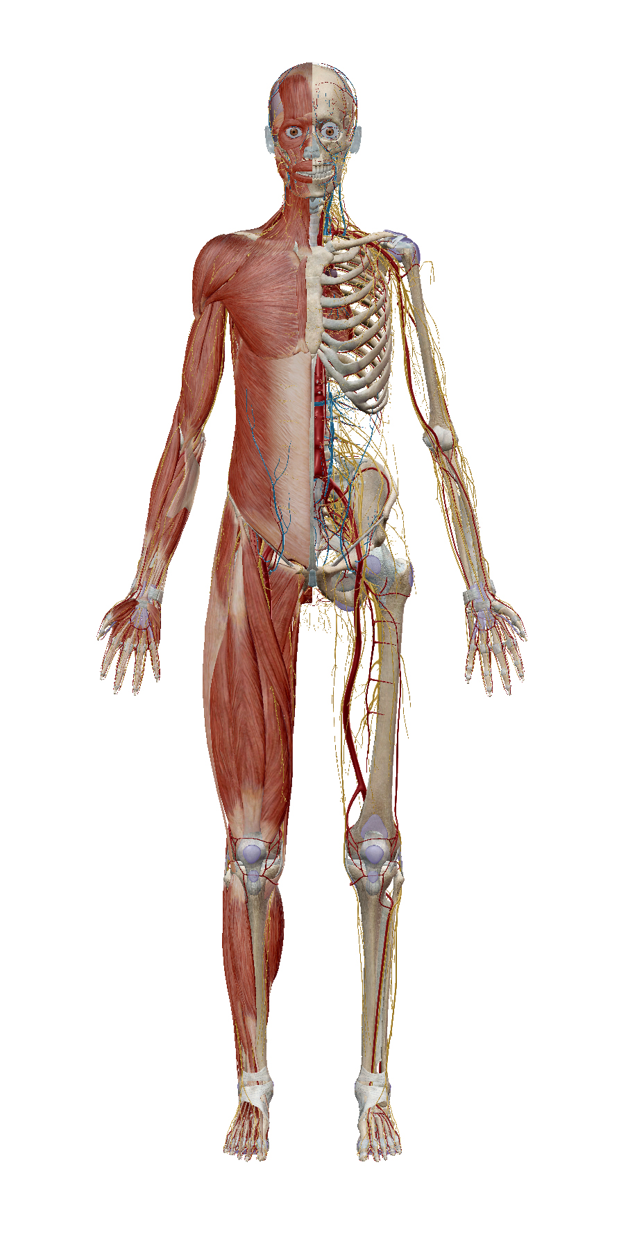
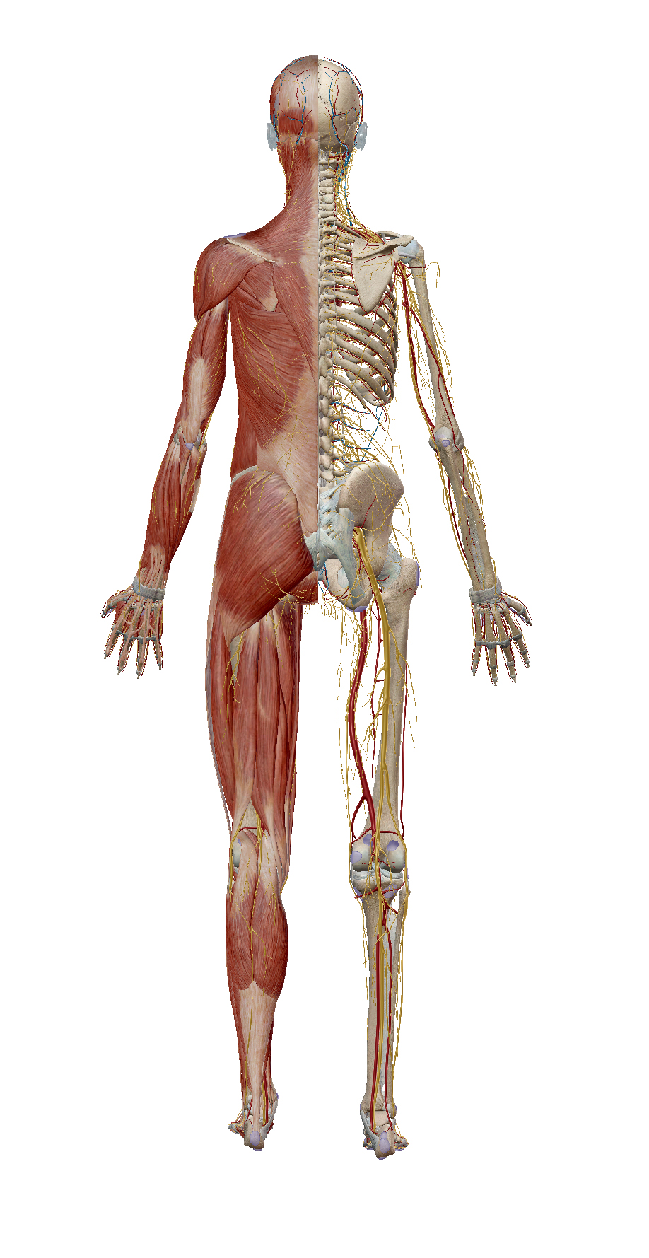
They are carried out through the musculoskeletal system. The musculoskeletal system includes the bones connected to each other in the skeleton and the muscles.
The passive part of the musculoskeletal system is the skeleton and the active part is the muscles.
The skeleton and muscles ensure the movement of the body in space, perform movements during work and speech, overcome the force of gravity.
Except as organs of movement some bones perform a supporting and protective function. Form cavities in which important internal organs are located.
In the cranial cavity, for example, is the brain, and in the thorax is the heart and lungs.
Skeletal muscles are attached to different parts of the skeleton and their contraction is controlled voluntarily. It is necessary for the performance of various movements and the movement of the body in space.
The heart muscle is involved in building the cavities of the heart, and although it is transversely striated, it has a number of properties that distinguish it from both smooth and skeletal muscle.
The muscle cell contains many fibers, myofibrils, running along its entire length.
The myofibrils themselves are made up of even thinner fibers called myofilaments, which are made up of protein molecules and are of several types. Myosin and actin myofilaments are particularly characteristic.
They are not indiscriminately piled up, but follow a strictly defined order. Muscle cells are grouped into individual muscle bundles, which in turn form the entire muscle.
Basics about muscles and tendons
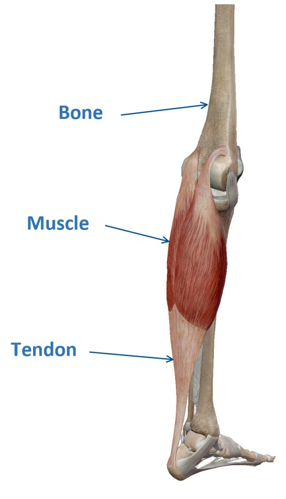
Every muscle has a beginning and a catching place. At both ends, muscle tissue attaches to the corresponding bones, most often through tendons.
They are made up of dense connective tissue and contain a scarce amount of blood vessels. Some tendons are covered with a two-layer sheath of fibrous connective tissue, the vaginal tendon.
The two contacting sheets of this shell are smooth and the small space between them is filled with liquid.
This greatly facilitates the movement of the tendon, as its inner leaf adheres tightly to it, and the outer connects with the surrounding tissues. Some muscles do not attach to the bones, but to the surrounding soft tissues.
Such are the facial muscles. When they are shortened, the mutual arrangement of the tissues on the face changes and the corresponding facial expressions are obtained.
Some details about muscles
There are about 650 different muscles in the human body. They can be classified according to different criteria:
- In Shape
- Location
- Function
- Fiber direction
In shape – the muscles can be long, short and wide. The long and short muscles are mostly spindle-shaped and have a body and two ends – a head at the beginning and a tail at the capture site. Some muscles have more than one head. Such are the biceps and triceps muscles of the armpit, the quadriceps muscle of the thigh.
Some muscles have more than one tail or several tendons. The common flexor of the fingers (and also of the toes) for example have up to four tendons. Thus, when only one muscle is contracted, its action extends to several fingers simultaneously.
Some muscles originate from different parts of the body and subsequently merge into a single muscle separated by tendon strips.
In such cases there is an alternation of passive and active elements and in practice the muscle is divided into several sections. An example is the rectus abdominis.
The broad muscles do not have well-defined pointed edges and end in a dilated broad tendon. These muscles are mainly on the torso – some of the muscles of the back and abdominal wall.
- Folders
- Unfolders
- Adjusters
- Drains, etc.
When muscles are directly connected to the performance of a movement, they are called synergists. And the muscles involved in the opposite movement are antagonists.
There are several muscles in the human body that do not work on their own. They are organized in groups. Different movements load different muscle groups, and the other ones rest. Almost half of our weight is due to muscles. For your workouts in general muscles can be simplified as:
- Large muscles
- Small muscles
Voluntary muscles
Most of our movements, such as the movements of our arms and legs, are controlled by our thoughts. The actions of the muscles are controlled by the brain, which sends and receives signals from the nervous system. The muscles involved in these movements are driven consciously and are called voluntary.
Involuntary muscles
There is another type of muscle in which the movements are performed automatically by the brain (movements of the heart, diaphragm, intestines, breathing, etc.). These muscles are called involuntary. One does not need to think to cause the work of these organs. For example, the heart beats at 60 to 80 beats per minute without us thinking about it.
Some details about bones
The human skeletal system is made up of 206 parts – the bones themselves. Depending on where they are located and the function they perform, bones have different shapes and sizes. In shape and structure, bones are of three main types: long, short and flat.
The long bones are located in the limbs and provide fast movements with a wide range. They look like levers driven by skeletal muscles caught in them. In long bones, there is a middle part called the body and two ends. The body resembles a cylinder and is made up of dense bone. Inside is a canal.
Short bones form complexes that provide strength, mobility and flexibility. They make up the spine, wrist and foot.
Flat bones are those of the skull, sternum, ribs, shoulder blade and upper pelvis. They form cavities in which important organs are housed. The flat bones consist of two plates of dense bone and a spongy bone substance with red bone marrow located between them.
Skeleton
In the process of its historical development, as a result of upright walking and human activity, changes have occurred in the skeletal system. The vertebral column, unlike the vertebral column of animals, has taken the form of a stretched double S with 4 curves and is a flexible spring column.
The stronger development of the brain and the weaker development of the jaws have led to changes in the relationship between the brain and the facial skull.
The thorax is made up of the sternum and 12 pairs of ribs. The sternum is located in front and has the shape of an elongated plate. The ribs have the appearance of narrow curved plates, which at the anterior end turn into cartilage. Each rib is movably connected at one end to the respective thoracic vertebrae, and its other end is connected to the sternum by cartilage.
The capture site for the spine is higher than the capture site for the sternum and thus allows the ribs to be raised and relaxed during respiratory movements. The last two pairs of ribs do not connect to the sternum, remain free and are called floating ribs.
Upper limb
The skeleton of the upper limb is made up of a shoulder girdle and a free upper limb. The shoulder girdle consists of a clavicle and a shoulder blade. The clavicle is a long bone in the shape of a stretched S. Through joints it is caught with one end for the sternum and with the other for the shoulder blade.
The shoulder blade has the shape of a triangular plate, for which many muscles are caught. Some of them attach it to the chest wall. The pit of the shoulder joint is located on the shoulder blade. The free upper limb consists of an arm, a forearm, a wrist and a hand.
The arm is formed by the humerus, which with its upper convex end connects with the fossa of the shoulder blade and forms the shoulder joint.
This is the most mobile joint: here the upper limb is brought forward, backward, sideways and retracted to the torso, as well as its rotation along the longitudinal axis.
At its lower end, through the joint, the humerus articulates with the bones of the forearm and forms the elbow joint. There are two forearm bones.
Lower limb
The skeleton of the lower limb is made up of a girdle and a free lower limb. The girdle of the lower limb is called the pelvis. It consists of two pelvic bones, the sacrum and the coccyx.
The posterior pelvic bones are connected to the sacrum in the semi-mobile pelvic joint, and the front are fixedly connected to the cartilage plate. The massive bone ring of the pelvis is the support of the spine. Through it, the weight of the body is transmitted to the lower limbs.
The pelvis is the bony protection of a number of internal organs and a place to capture many muscles. On the outside of the pelvis is a deep articular fossa to connect to the femur.
The free lower limb is made up of the thigh, lower leg and sole. The thigh is formed by the femur – the longest and strongest bone in the human skeleton. At its upper end is a large head, which is articulated with the pit of the pelvis in the hip joint.
It is similar to the shoulder joint, but the movements are more limited at the expense of greater joint strength. The lower leg consists of two parallel long bones: a large tibia located on the inside and a small tibia connected immovably.
The upper end of the tibia is involved in the formation of the knee joint. It articulates the femur, lid and tibia. It is strengthened by strong connections, two of which cross the joint cavity. In front is a small bone-cap.
In the knee joint, the lower leg is folded and unfolded. The unfolding of the 180 is prevented by the ligaments of the joint, not by the cap, which serves to capture the quadriceps muscle of the thigh.

Leave a comment